Saturday, November 28, 2015

Instantaneous 3D bone pose

In order to describe the pose of the bones in a ridged body we use a reference frame with local coordinates using bone points and invariant time. This reference frame is called the instantaneous 3d bone pose.
The most complicated aspect of the pose is the mathematical description. One must describe the position and orientation of the bone (local reference plane) with respect to a global reference plane.
In the case of the 3D bone pose there are six scaler quantities, each on a sampled instant of time, a position vector, orientation vector, and a orientation matrix. The orientation matrix of the local frame with respect to the global one is defined by a 1st column the xl axis versor (Versor is a templated class that holds a unit quaternion. The difference between versors and quaternions is that quaternions can represent rotations and scale changes while versors are limited to rotations) components, the second column, the yl axis versor components, and the third column, the zl axis versor components. The three components stated above are the cosines of the angles between each versor and the XYZ global axes. Here is a numerical description of the pose vs time (3-D case).

Forces Transmitted by Muscles-Tendons-Ligaments-Bones

Movement occurs through the coordinated working of bones, tendons and ligaments. All three pertinent parts of the human body work together in response to neurological signals, and if there is any disease or condition that interrupts the nerves' signals or if there is any injury to any of these structures, movement can be hindered. In order to gather a fuller understanding lets recount what tendons, bones and ligaments are. Tendons are tough bands of connective tissue found in the joints. They connect muscles to bones. Each muscle has tendons attached at each end. Tendons are designed to only stretch a small amount. Their job is to transmit force between the bones and the muscles. For example, when the biceps muscle on the front top of the arm contracts, the tendon attached to the biceps muscle and elbow bone helps the muscle to pull on the elbow bones so the joint can bend. Ligaments are made of the same material as tendons. Ligaments connect the bones to each other, and are designed to help stabilize the joints and provide a structure for the bones. Since they have limited stretching ability, they limit how far a joint moves to help protect against injury. As the elbow joint bends, the ligaments stabilize the elbow bones so the arm can move with control.

Muscles in the body contract or shorten when they receive nerve signals initiated by the brain. There are three types of muscles including, skeletal muscles, which can be voluntarily controlled, involuntary smooth muscles, such as those that control breathing, digestion and other functions, and involuntary cardiac muscles, which control the function of the heart. Skeletal muscles travel across the length of joints and stretch between the bones.
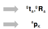
Forces transmitted by muscles can be illustrated using Internal Load Modeling. Muscles, ligaments and tendons are treated as ropes, thus there is no 3D modeling for their forces. Internal Load Modeling does not take into account any interaction with surrounding muscles and bony structures. For each body segment of interest, the following quantities are estimated: the position vector and orientation matrix relative to both the laboratory frame (g)  and the anatomical frame (a), plus the local position vector of the intersegmental loads which is represented by reduction point K.

The left most diagram below depicts forces acting outward on the knee structure, and to the right shows the forces acting downward on the lower half of the leg.


Kinematic quantities and Inertia parameters are taken into consideration when using Internal Load Modeling as well as the forces shown above. For each body segment of interest, the following quantities are estimated in addition to the ones stated above... Mass is taken into consideration represented by m and K is no longer an arbitrary point.  The local position vector is represented by the notation CM (note in the equation CM is broken up into sub vectors) and the principal axes of inertia by I. The orientation matrix a is also considered as well as moments of inertia. All such things are coupled together to estimate Intersegmental Force (the force between two segments,) as represented in the equation below.


Muscular Mechanical Work/Power & Joint Energetics

Muscle mechanical work is an important biomechanical quantity in human movement. Power is defined as the rate of work or the rate of energy flow. Two power measures can be obtained from the joint kinetics: the joint power and the muscle power. The joint power is the scalar product of the net joint force and the joint velocity (P(j)=F*V)where P(j) = the joint power, F = the net joint force, and v = the velocity of the joint. Precisely speaking, the joint power is the rate of energy transfer through the joint caused by the linear motion of the joint.
where FAK/FT = the net joint force acting on the foot at the ankle, FAK/SH = the net joint force acting on the shank at the ankle, and vAK = the ankle velocity. [3] shows that both the foot and leg joint powers are of the same magnitude with an opposite sign at the ankle. This suggests that the ankle joint only transfers energy from foot to the leg and vice versa. Between the two segments forming a joint, one segment always gains energy at the rate the other loses its energy and vice versa.
Muscle power on the other hand is a scaler product of joint torque and the segment's angular velocity (P(m)=T*w)where P(M) = the muscle power, T = the joint torque, and w = the angular velocity. When looking at muscle powers of the foot and the leg at the ankle we can conclude...
where TAK/FT = the ankle joint torque acting on the foot, TAK/SH = the ankle joint torque acting on the lrg, wFT = the angular velocity of the foot, and wSH = the angular velocity of the leg. It appears that there is no apparent relationship between the muscle powers of the foot and leg since the angular velocities of the foot and leg can be very different.At the muscle, two things happen: (1) the muscle transfers energy from one segment it attaches to to the other, and (2) the muscle does work through contraction. The energy transfer happens from one segment to the other and the net change in the energy in the two segments due to the energy transfer must be 0. On the other hand, an individual muscle can either add (positive work) or drain (negative work) energy to or from both segments at the same time by doing work. The angular momentum is shown in figure 2.

Selective Glossary

energetics the branch of science dealing with the properties of energy and the way in which it is redistributed in physical, chemical, or biological processes. Thoracic Centra Middle segment of the vertebral column, there are 12 Thoracic vertebrae between the cervical and the lumbar vertebrae sagittal suture The dense fibrous central tissue between the two parietal bones of the scul, begin to close at age 29 and is fully closed by the age of 35. distal segment The portion of the joint furthest from the body Proximal segment opposite of distal segment, the segment nearest to the center of the body. Cartesian coordinates system is a coordinate system that specifies each point uniquely in a plane by a pair of numerical coordinates, which are the signed distances to the point from two fixed perpendicular directed lines, measured in the same unit of length, synovial joint A synovial joint, also known as diarthrosis, joins bones with a fibrous joint capsule that is continuous with the periosteum of the joined bones, constitutes the outer boundary of a synovial cavity, and surrounds the bones' articulating surfaces. The synovial (or joint) cavity is filled with synovial fluid. Pin Joints a mechanical joint that will transmit axial load but will not transmit torque

Reflection on the Research

I began my project believing I would be able to fit all my information into one stop motion animation. It was a struggle for me trying to figure out how I would be able to pack all of my research and hard work into one video less than 5 minutes long. However I came to the harsh realization that I just would not be able to do so. I discussed with my teacher Mr. Lyke  about adding a website link to my video where I would display all of my research. I had already decided on researching Biomechanics and the human body, and had a basic layout for what I wanted to discuss in my project. I did basic research and had an understanding about what Kinematics and Kinetics was prior to the construction of my website. Once I began constructing my website I was able to expand upon a majority of my concepts I was addressing in comparison to when I was only doing a video. When conducting my research I did a majority through the search engine google scholar. The most challenging part of my project however did not come in finding enough research and understanding the content but rather technological difficulties. The difficulties first began in trying to construct a stop motion and having glitches in the slide timing. Then came constructing a website. I tried multiple system and could not find any easy to use builders. In the end I decided to use Blogger and set it up as it was a website, with tabs and non sequential order. I added a link to my video on the website and a link to website on my you tube account. All in All I am fairly happy with the work I did and with my website. I throughly enjoyed connecting human movement to physics and found a lot of fascinating websites and informative research.

Works Cited

Works Cited

A., and Advanced Technologies For Neuro-Motor Assessment And Rehabilitati. ADVANCED TECHNOLOGIES FOR NEURO-MOTOR ASSESSMENT AND REHABILITATION BIOMECHANICS OF HUMAN MOVEMENT (n.d.): n. pag. Web.
Bartlett, Roger. "Introduction to Sports Biomechanics." (1997): n. pag. Web.
"Biomechanical Analysis of Fundamental Human Movements - Arthur Chapman." Human-kinetics. N.p., n.d. Web. 10 Dec. 2015.

"MVS 330: Biomechanics of Human Movement." MVS 330: Biomechanics of Human Movement. N.p., n.d. Web. 10 Dec. 2015.